Discover powerful AI-based tools to enhance your digital pathology workflows.
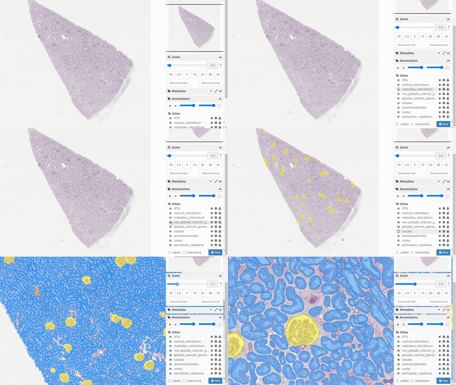
Analyze kidney WSIs by segmenting six compartments, leveraging Detectron2-based AI models.
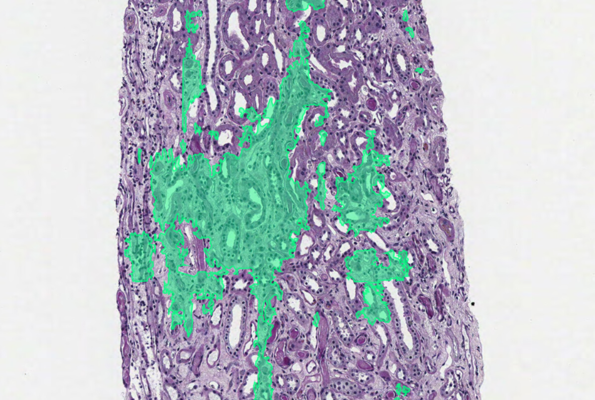
Automate the detection and quantification of Interstitial Fibrosis, Tubular Atrophy, and Glomerulosclerosis.
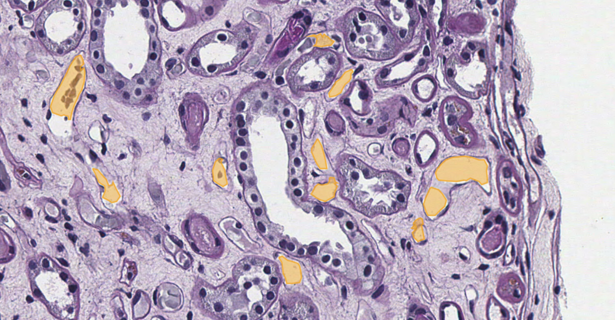
Quantify PTC density, size, and shape to gain insights into microvascular changes.
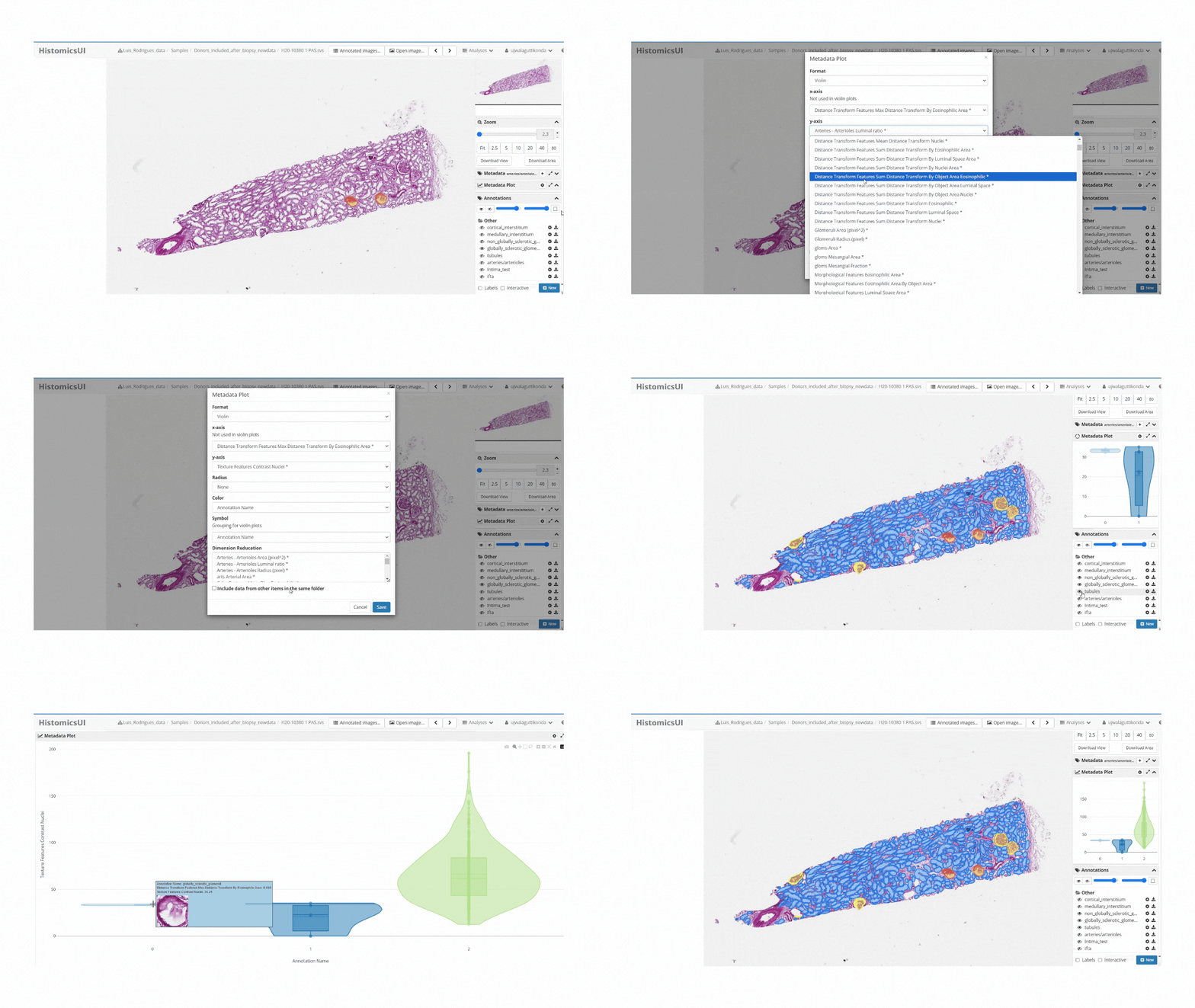
Extract key structural and morphological features for advanced clinical insights.
Leverage our Detectron2-based model to analyze kidney WSIs by identifying and segmenting six compartments: cortical interstitium, medullary interstitium, non-sclerotic glomerulus, sclerotic glomerulus, tubule, and artery/arteriole.
Back to PluginsThe IFTA Segmentation tool automates the detection and quantification of Interstitial Fibrosis, Tubular Atrophy (IFTA), and Glomerulosclerosis in kidney tissue. This tool enables efficient and accurate segmentation from renal biopsy images.
Back to PluginsPeritubular Capillary (PTC) segmentation quantifies PTC characteristics such as density, size, and shape in kidney biopsies, providing insights into microvascular changes and disease progression.
Back to PluginsOur feature extraction module identifies and segments key kidney structures (glomeruli, tubules, arteries) using deep learning. It then quantifies key metrics (diameters, areas, densities) and correlates these with clinical parameters like age, serum creatinine, and eGFR for better understanding of kidney morphology and disease implications.
Back to Plugins